-
Phone
86-371-61315851
-
Address
Room 806, Floor 8, Wanda Center, Jinshui District, Zhengzhou, Henan, China
-
E-mail
LatestProducts


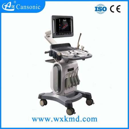
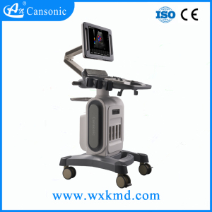
Diagnostic System Full Digital Trolley Color Doppler Ultrasound Scanner
China Diagnostic System Full Digital Color Doppler Ultrasound Scanner (YJ-U60), Find details about China Color Doppler, Ultrasound Scanner from Diagnostic System Full Digital Color Doppler Ultrasound Scanner (YJ-U60) - Henan Forever Medical Co., Ltd.
Description
Basic Info
- Model NO.: YJ-U60
- Customized: Customized
- Scale: Large
- Focus: Continuous Dynamic Focus
- Color: Black
- Zoom: 10 Steps
- Atmospheric Pressure: 700hpa to 1060hpa
- Probe Transducer Types: 3.5 MHz Convex Probe
- Transport Package: Standard Importing Cartons
- Origin: China
- Medical Device Regulatory Type: Type 2
- Certification: SGS, CE, ISO13485
- Model: Yj-U60
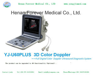
- Name: 3D Color Doppler
- Frequency: 50Hz+-1Hz
- Power: AC 100V to 240V
- Keyboard: Light Floating International Standard
- Trademark: forermed
- Specification: 510 mm X 500 mm X 330mm
- HS Code: 9018129100
Product Description
Best Protable Medical Equipment Ultrasound Color Doppler

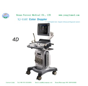
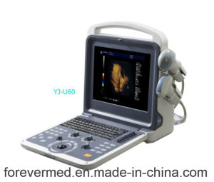
YJ-U60 3D Color Doppler Full Digital Color Doppler Ultrasound Diagnostic System
12' high-resolution LED monitor, delicate appearance,
Exquisite image quality and excellent stability,
Protable ultrasound diagnostic system.
Applications
Abdomen, Obstetrics, Gynecology, Pediatrics, Small parts,
Artery, Superficial organ, Orthopedic, Cardiology, etc.
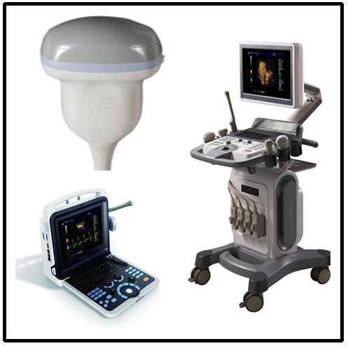
Appearance
4D volume probe (2.0/ 3.0/ 3.5/ 4.0/ 5.5Mhz)
Function
Connectivity
Standard Accessories

For more Hospital machine detailed information, please feel free to contact with our sales team.We are always here to service for you!
Contact Person: Ms. Fiona
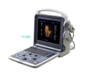
YJ-U60 3D Color Doppler Full Digital Color Doppler Ultrasound Diagnostic System
12' high-resolution LED monitor, delicate appearance,
Exquisite image quality and excellent stability,
Protable ultrasound diagnostic system.
Applications
Abdomen, Obstetrics, Gynecology, Pediatrics, Small parts,
Artery, Superficial organ, Orthopedic, Cardiology, etc.
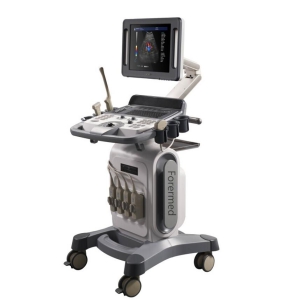
Appearance
- Smart, compact and clamshell design
- 12 inch LED monitor
- Backlit operation panel, 8TGC
- Floating keyboard
- Two active probe connectors
- Two probe holders
|
Probe |
5 Steps Multi-frequency |
| 3.5Mhz convex probe | (2.0/ 3.0/ 3.5/ 4.0/ 5.5Mhz) |
| 6.5Mhz transvaginal probe | (5.0/ 6.0/ 6.5/ 7.5/ 9.0Mhz) |
| 7.5Mhz linear probe | (6.0/ 6.5/ 7.5/ 10.0/ 12.0Mhz) |
| 3.5Mhz micro convex probe | (2.0/ 2.5/ 3.5/4.5/ 5.0Mhz) |
| 3.5Mhz phased array probe | (2.1/ 3.0/ 3.5/ 4.0/ 5.0Mhz) |
Function
- Auto Image Optimization
- Tissue Harmonic imaging
- iClear (Speckle Noise Reduction)
- iBeam (Spatial Compound Image)
- iZoom
- PIHI (Pulse-Inverse Harmonics Imaging)
- SA (Synthetic Aperture ultrasonic Imaging)
- Panoramic Image (Option)
- Trapezoid Image (Option)
- Continuous Wave Doppler(Option)
- B, B|B, 4B, B|M,M,B|D,PW,B|PW, CF
- Duplex/Triplex mode
- CW (option)
- 4D mode (option)
Zoom
Cine‐memory
- Real time zooming
- Selectable zooming position
- Continuous dynamic focus
- 1~16 selectable transmit focus
- Acoustic lens focus
- 1, 2, 3, 4 focus
Cine‐memory
- B‐mode M‐mode
- SSD (Solid State Disk) 64G
Imaging Processing
B mode
- Depth: 1.6~30cm
- Noise reduction: 0-6
- Gain: 0~100%
- Gain: 0~100%
- Gain: 0~100%
- Sampling volume: 0.5-48mm
Measurement & Calculation
Measurement
B mode (General)
Abdomen
B mode
- 8‐step TGC slide pots
- Depth: 1.6~30cm
- Frequency: 5 steps
- Dynamic range adjustable: 0~150dB
- Edge enhancement:0~7
- Persistence:0~7
- Chroma:0~6
- Grayscale:0~16
- Noise reduction: 0-6
- iclear: off, 1, 2, 3, 4
- Gain: 0~100%
- Sweep speed: 4 steps
- Chroma:0~6
- Gain: 0~100%
- Pulse wave
- Wall filter: 4 steps
- Color Maps: 0~7
- Package size: 8~15
- Color persistence: 0~7
- Threshold: 0-3
- Base line: 0-6
- Line density: Low and high
- Spatial filter: 0-3
- Gain: 0~100%
- Frequency: 5 steps
- Pseudo color:0~6
- PRFd:1.0~6KHz
- Basic line: 7 steps
- Wall filter: 7 steps
- Sampling volume: 0.5-48mm
Measurement & Calculation
Measurement
B mode (General)
- Distance
- Trace Length
- Ellipse (area)
- Trace(area)
- Angle
- Volume
- HR (heart rate)
- Distance
- Velocity
- Time
Abdomen
- Liver
- Gallbladder
- Pancreas
- Spleen
- Kidney
- Ureter
- Bladder
- After the urine bladder
- Prostate
- Uterus
- Cervix
- Ovary
- Follicle
Early Obstetrics
Musculoskeletal- GS
- BPD
- CRL
- NT
- BPD
- HC
- AC
- OFD
- FL
- TAD
- Thyroid
- Testes
- Hip
- Intima
- Artery
- Distance
- Angle
- Volume
- RVWd
- LVDd
- RVDd
- LVPWd
- RVWs
- LVDs
- RVDs
- LVPWs
- RV/LV
- AO
Connectivity
- Video out port
- DVI out port
- VGA out port
- 2 USB port
- DICOM 3.0
- Gross dimension: 510 mm X 500 mm X 330mm
- Net dimension: 330mm X 150 mm X 380mm
- Gross weight -- 12 kg
- Net weight -- 7 kg
- Voltage: AC 100V to 240V±10%
- Frequency: 50Hz±1Hz
- Rated Power: 250VA
- Ambient temperature: 0ºCto +40ºC
- Relative humidity: 38% to 85%
- Atmospheric Pressure: 700hPa to 1060hPa
Standard Accessories
- Power Cable
- Operation Manual
- Fuse
- System Recovery USB
- Built in Li-ion battery
- B/W or color Video printer
- LaserJet or inkjet printer
- Trolley
- Aluminum case
- Biopsy guide
For more Hospital machine detailed information, please feel free to contact with our sales team.We are always here to service for you!
Contact Person: Ms. Fiona